Our researchers conduct experiments on the following equipment:
Optical microscopy
Digital optical 3D microscope Hirox KH-7700 (HIROX, Japan)
Scaning probe microscopy
Atomic force microscope Ntegra Prima (NT-MDT BV, Netherlands)
Sample preparation
Cryo ultramicrotome Leica EM Ultracut7 (Leica, Austria)
Spincoater WS-400BZ-6NPP/A1/AR1 (Laurell, UK)
Vacuum thermochamber ТИРСА-РС (Актан Вакуум, Russia)
Properties of thin films
Spectral ellipsometric complex ELLIPS-1891 SAG (Russia)
Mechanical testing
Universal testing machine Testometric FS100 CT (Testometric, UK)
Dynamic Mechanical Analyzer DMA/SDTA861e (METTLER TOLEDO, Switzerland)
Biaxial testing machine Zwick (Zwick/Roell, Germany)
Digital optical 3D microscope Hirox KH-7700 (HIROX, Japan)
Obtaining digital images of micro-objects and measurements in 3D.
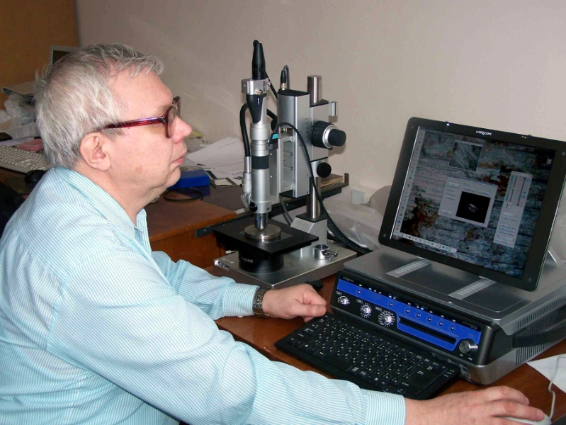
- 3D measurements of object’s surface.
- Optical zoom up to 7000x.
- Synthesis and comparison of images.
- Video recording (30 fps).
- 3D-view (360°) with adjustable viewing angle from 25° to 55°.
- Measurements at variable angles of illumination, with differential interference contrast, dark and bright field, polarized light.
- 3D-reconstruction of deep features of the surface using multifocus capabilities.
Atomic force microscope Ntegra Prima (NT-MDT BV, Netherlands)
The study of material surfaces at the micro- nano-scale.


- Scan area: up to 100х100 microns.
- Max resolution in xy plane: ~1…10 nm (depends on the probes).
- Max vertical resolution (z): < 0.1 nm.
- Measurements in dry room conditions.
- Nanolithography.
- Scanning tunelling microscopy.
- Mapping of electrical conductivity.
- Nanomechanical mapping.
Cryo ultramicrotome Leica EM Ultra Cut 7 (Leica, Austria)
Obtaining ultrathin sections and fine flat surfaces. Preparation of sections for transmission electron microscopy (located in Perm University).
Convenient to prepare the surface for the probe / electronic / spectroscopic analysis of the surface (filled) polymers, biological tissues, including hard materials like metals and bones.
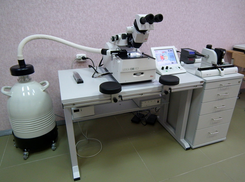
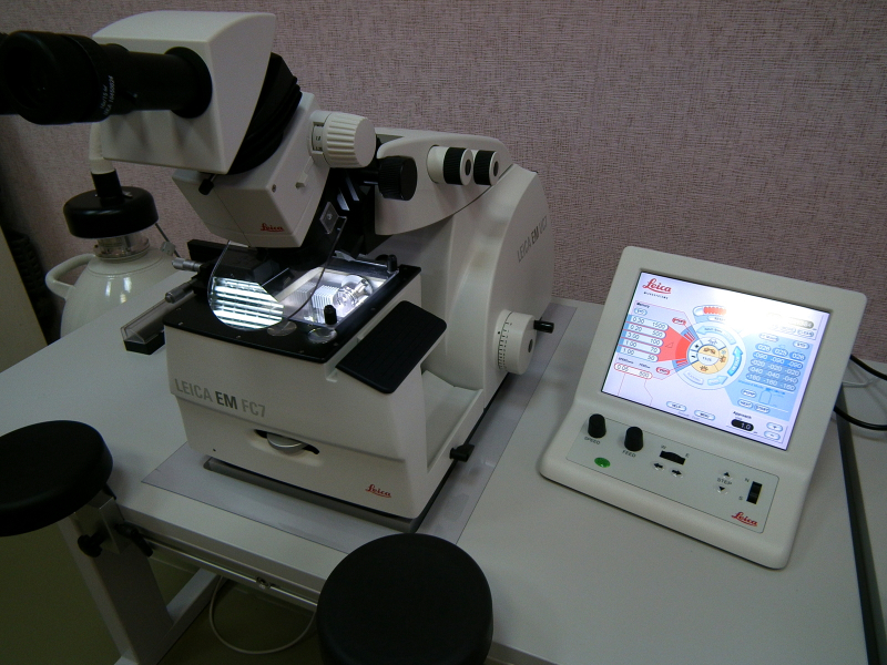
- Equipped with cryo-chamber Leica FC7.
- Minimal section thickness ~ 10 nm.
- Three regimes of low-temperature sectioning: normal, wet and dry cutting.
- Equpped with glass or diamonds knifes.
Spincoater WS-400BZ-6NPP/A1/AR1 (Laurell, UK)
Deposition of thin films from solution onto a smooth surface by centrifugation.
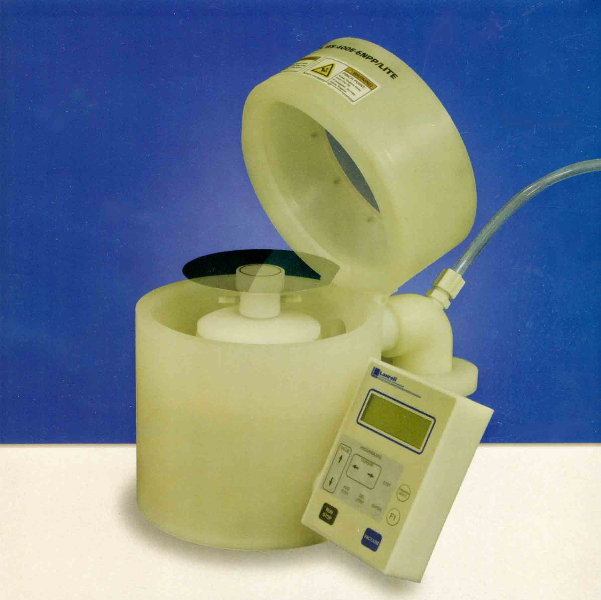
- Internal chamber size: 216 mm.
- Diameter of the substrate: 150 mm.
- Spinnig velocity: 100-8000 r/min.
- Vacuum pad, pump and drainage tank.
Vacuum termochamber Тирса-РС (Актан Вакуум, Russia)
Drying in vacuum.

- Thermocontroller: multistep heating up to 250C.
- Max. vacuum < 0.002 Torr.
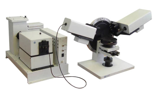
Spectral ellipsometric complex ELLIPS-1891 SAG (Russia)
The measurement of the spectral dependences of the optical parameters of the surface structures, determination of optical and structural properties of materials and thicknesses of thin-film layers.
The spectral range – 350…1100 nm.
Universal testing machine Testometric FS100 CT (Testometric, UK)
- Maximal force – 100 kN.
- Maximal dispacement – 1200 mm.
- Velocity: 0.001 … 500 mm/min.
- Additonal force sensors.
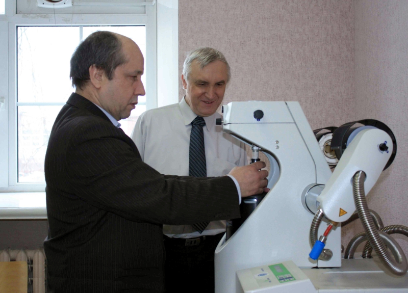
Dynanic modulus analyzer DMA/SDTA861e (METTLER TOLEDO, Switzerland)
Dynamic mechanical analysis of the materials (located in Perm Iniversity).
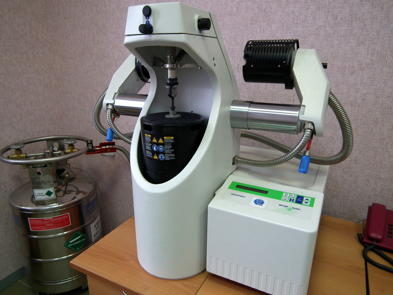
- Length of the sample – 10.5 mm.
- Frequency range of harmonic oscillations: 0.002 – 200 Hz.
- Force amplitude range: 0.005 – 18 N.
- Deformation amplitude range: up to 1600 microns.
- Temperature range: -150°C – +500°C.
Biaxial testing machine Zwick (Zwick/Roell, Germany)
4-directional testing machine with mutually perpendicular axes in one plane load. Biaxial test specimens in tension and compression (located at Perm University).
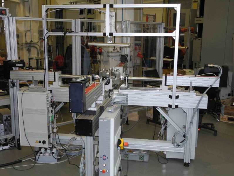
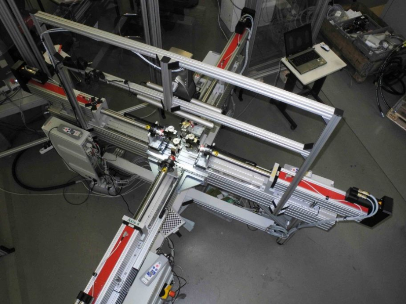
- Max loading ± 2 kN.
- Total displacement of one axis – 800 mm.
- Total velocity of one axis: 0.001 mm/min – 15000 mm/min.
- Equipped with video extensometer videoXtens Array.
Last updated: 16-02-2021